Management of Chronic Osteoarthritis in Dogs
Introduction
 Osteoarthritis (OA) is a degenerative condition of tissues in and/or around a joint. It is not only the bones that are affected but also the cartilage covering the ends of the bones, the capsule that surrounds the joint and the ligaments that support the joint. It is not the same as Rheumatoid arthritis in people and although this does occur in dogs it is quite rare. OA in dogs is a disease process that results from damage to the joint. The damage may be a result of a poorly fitting joint (e.g. in hip or elbow dysplasia), a consequence of joint instability (e.g. following rupture of a ligament) or else may follow direct trauma to the joint surface (e.g. a fracture involving the joint itself). Osteoarthritis is not a curable condition but it can often be successfully managed in one of several ways.
In some dogs with OA the signs may take the form of intermittent lameness following over activity and such “flare-ups” are generally best managed by restriction of exercise +/- a short course of non-steroidal anti-inflammatory drugs (NSAIDs, one type of pain killer), which may be prescribed by your veterinary surgeon. However, in dogs with established / persistent clinical signs relating to OA, management has to adopt a more long-term strategy and it is this situation that will be considered in this information sheet.
What signs are seen in clinical OA?
A dog showing clinical signs of OA may show stiffness on rising, a shuffling forelimb or hindlimb gait, reluctance to exercise, lameness at exercise, difficulty climbing into the car / up steps (hindlimbs), difficulty coming down steps / slopes (forelimbs). However, other problems can mimic OA, for example degenerative conditions of the lower back or ligament problems in both knees can cause identical signs “from the outside” as hip OA. Only after a clinical examination by a veterinary surgeon and radiographic (X-ray) confirmation of their suspicions should a diagnosis of OA be made. However, X-rays alone should never be used to make a diagnosis since what they look like bears no direct relationship with how painful a joint is, or indeed whether it is painful at all.
How is OA Diagnosed?
OA is usually diagnosed from the clinical signs being shown by a dog together with a joint showing thickening and reduced movement with pain on manipulation. X-rays will usually be required to rule out other causes of pain and should show typical signs of OA affecting the joint – generally production of bone around the joint edges. It must always be remembered that what is seen on X-ray and what signs a dog is showing are very poorly related, i.e. how severe the OA is should be measured by the clinical signs and NOT what the X-rays look like. Many dogs with horrendous looking X-rays are actually not lame.


Normal elbow – 6 year old Yorkie OA elbow in 5 year old Labrador
A combination of clinical signs and X-ray changes are usually all that is required to make a diagnosis but in some cases it is necessary to take some fluid from the joint and have laboratory analysis carried out to ensure there is no infection present that would explain a recent deterioration in the lameness.
How can the response to treatment be monitored?
If any treatment is to be given as a long-term measure it is important that its effectiveness is monitored and the owner is in the best position to observe this. However, these observations need to be recorded so that patterns can be recognised which may help to improve management by “fine tuning” what is being done. Different dogs show different “primary” signs to their owners and so what is to be monitored varies between dogs. Essentially, each factor should be scored out of ten (0=normal; 10 = very bad) and any number of factors (lameness, stiffness, exercise tolerance etc.) can be included – although however many are chosen must be included each time a score is given. The scores given to the factors are then totalled to give a measure of overall disability and this number is recorded in table or graph form. To begin with a score may be given each day but with time it may be found that once each week will suffice. Some pharmaceutical companies produce “pain calculators” and record charts to help measure and monitor a dog’s disability and it may be worth asking your veterinary surgeon about these.
What options are available to try and control the signs?
The options available can be divided into three broad categories: lifestyle changes; non-surgical measures; surgical measures.
1) LIFESTYLE
Within this group are two main considerations and these should be looked at first for any patient showing signs of OA:
Activity levels – The amount of exercise allowed may have to be reduced to allow a dog to cope. However, a sensible regime should be adopted since inadequate movement of an arthritic joint will allow stiffness to increase. Lead exercise is less likely to aggravate an OA joint than off lead exercise. Therefore, in general, the dog should be taken for regular lead exercise each day (not minimal during the week and excesses at the weekend) with the distance being dictated by what the dog can manage without deterioration in the signs being monitored. Hydrotherapy can be a very useful way of maintaining strength in the limbs without traumatising the joints and swimming sessions twice weekly for 4 or 5 weeks can produce improvements in activity levels that last for several months.
If the activity levels that can be attained are insufficient to allow a good quality of life or acceptable function then other measures have to be turned to.
Obesity – Being overweight will make any dog less able to cope with an OA joint and so will tend to make the clinical signs worse. A reduction in bodyweight is often all that is required to allow a dog to return to normal levels of activity. When dieting a dog it has been shown that prescription weight loss diets are better than so-called “starvation diets”. This is because giving a dog less food causes it to lose both fat and muscle in the same proportion, whereas the prescription diets are formulated so as to retain muscle and lose more fat.
2) NON-SURGICAL MEASURES
These are aimed at controlling the pain and thus improving function. Although some products may claim to have an effect on the progression of the OA itself there is no scientific evidence to justify any such claim in the dog. It is probably best to introduce any of the following one at a time so that the value of each can be monitored.
“Conventional” medications (mainly Non-Steroidal Anti-Inflammatory Drugs – NSAIDs) – One product that is not an NSAID, but which can be useful in long-term control of disability resulting from OA, is Cartrophen-Vet. The way in which it achieves this is poorly understood but a course of four injections, given a week apart, may lead to significant improvement that lasts for several months, after which the course can be repeated. As already mentioned above NSAIDs can be used in acute “flare ups” but they can also be used as a long-term strategy. Two (Metacam and Rimadyl) are licensed for long-term use in the dog and so these are preferred to other NSAIDs. However, certain drugs appear to work better in some dogs than others and it may be necessary to find a suitable product by “trial and error”. Some of the human preparations are not suitable for dogs and cause severe (possibly life threatening) reactions. Therefore, your own veterinary surgeon should be consulted before any medication is given to your dog and his/her advice heeded.
The NSAID chosen should be effective in reducing the signs of OA, tolerated well by the dog (no side-effects such as vomiting or diarrhoea), preferably licensed for long-term use and easily administered. Although cost is a factor, by the time all other requirements are met the difference in cost is likely to be negligible. When used long-term it is important to establish the minimum amount of the drug that is required to give maximum benefit. To that end the following protocol should be adopted :-
- Give the recommended dose for one month and monitor the effect, if this is not satisfactory try another drug but if it is satisfactory then
- Reduce the dose by about 10% every 5-7 days (easiest with Metacam since it comes as a liquid) and monitor for any reduction in effect.
- It is important that the dose is not reduced more frequently as blood levels take time to change and settle.
- Keep a diary noting what dose was given and the “disability score”.
This should allow the “maintenance requirement” to be established. Owners may acquire a feel for any changes that need to be implemented according to circumstance, e.g. giving more drug when the weather is cold and wet, or on days when the dog is likely to be more active (weekend / visitors etc.). In the event of a “flare up” of signs the dose may be increased whilst exercise is reduced, and then as the signs come back under control the dose can be reduced again.
Nutraceuticals – These products are natural products, are given in food and may improve the signs in some dogs. They need to be given at the recommended dose and to decide upon how effective they are in a particular patient it is probably best to trial them for a month. There is little scientific evidence that they work in the dog, possibly because few relevant studies have been done. One study in dogs did suggest that extracts of green lipped muscle did help to reduce the clinical signs of OA. In humans there is some evidence to suggest that one particular component (glucosamine) is of value. Based on this it is probably best, if these products are to be used in dogs, that a preparation containing this component is used. Since the production of these preparations is poorly regulated it is probably best to use one made by a reputable manufacturer that has more to lose if the product was found to be of unsatisfactory quality.
Acupuncture – May provide an improvement in “disability score” that lasts for a prolonged period before the course of treatment needs to be repeated.
Homeopathy – This should not be disregarded as it may produce a favourable improvement in some cases and certainly will do no harm.
Others – Certain other products are often used because of anecdotal reports of their effects, including magnetic collars and cod liver oil. Although the latter might produce improvement care should be taken not to overdose with it since toxic levels in the dog are much lower than in humans, and as a rule it might be better to avoid the administration of such preparations.
3) PHYSIOTHERAPY
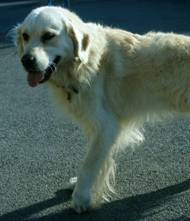
In recent years physiotherapy has become an available therapy for dogs. Osteoarthritis is just one condition that can be managed utilising physiotherapy, either alone or in combination with one or more of the measures outlined in this document. If this option is pursued then it is important to seek good quality physiotherapy. Unfortunately the term veterinary is not regulated for use only by Veterinary  Surgeons/Nurses and the term physiotherapist is not regulated for use only by those with qualifications in physiotherapy. Hence more or less anyone can call themselves a veterinary physiotherapist without necessarily having qualifications in either discipline! It is important, therefore, that you take advice from your Veterinary Surgeon as to where you should take your pet. Any such therapy is being undertaken under the supervision / authority of your Veterinary Surgeon (and they need to refer you to the person concerned) unless the veterinary physiotherapist is a Veterinary Surgeon themselves – when they are accountable and responsible for the therapy given. Weighbridge Referral Service has a close working relationship with the SMART clinic (picture courtesy of Smart Clinic) and their details can be obtained by way of their website www.smartvetwales.co.uk.
4) SURGICAL MEASURES
Surgery may offer a means of improving function by reducing or eliminating the pain from an arthritic joint. To avoid repetition below the general statement that surgery is generally only considered if the measures above prove inadequate is made here. The exact form of the surgery and the expected benefit varies depending on the joint involved but the following represents a summary :-

Shoulder – Rarely does OA cause clinical signs in this joint that cannot be controlled using non-surgical options. But if it does then arthrodesis (fusion) of the joint will eliminate the pain and have little effect on limb function.
Elbow – Debridement of any loose fragments from the joint by arthroscopy (key-hole surgery using the so-called “magic eye or camera”) or arthrotomy (conventional style surgery) may improve the level of disability, possibly bringing it back within the realms of control using the non-surgical measures listed above. Arthrodesis (fusion) of the elbow is possible and will eliminate pain from the joint. However, it does compromise function of the leg and it takes several months for a dog to adapt to not having a movable joint in the middle of the limb. Total elbow replacement – is becoming an accepted treatment for cases not responding to other management options. Improvements in design of the prosthesis are helping to reduce the complication rate. Because of the potential complications the treatment is only used in quite severe cases and the number performed annually in the UK is still (2012) probably measured in single figures or slightly more.


Arthroscopic (“magic” eye) view of OA in an elbow) Following surgery to fuse Elbow

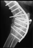
Carpus (wrist) – Arthrodesis (fusion) is very effective at eliminating pain from the joint and the carpus is probably the joint most often fused with the expectation of very good results. The commonest complications seen are infection and the plate used to “fix” the joint will need to be removed in about 25% of cases once fusion has been achieved, because of irritation under the skin.
(This picture on the lfet shows a carpus 4 months after surgery to fuse thejoint.)
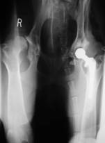
Hip – There are two surgical options for this joint and which is chosen is governed by the size of the dog and financial constraints. Total hip replacement is becoming a more frequent procedure in the dog and can result in a return to normal limb function. Initially, this procedure was only possible in larger breeds but with an increased range in size of the prosthetics available it is possible to perform such surgery in most patients. However, this is somewhat tempered by the better results seen using the next procedure to be outlined in the smaller patients compared with their larger counterparts. The alternative is to remove the ball of the hip joint and leave nature to form a so-called “false joint” (though this should not be confused with a hip replacement). The results of doing this are generally good in smaller patients but less so in the larger breeds. However, the use of good quality physiotherapy post-surgery can improve the end result enormously.


Hip after removing the ball of the joint Hip after total joint replacement
to form a “pseudoarthrosis”
Stifle (knee) – Debridement of any loose fragments from the joint by arthroscopy (key-hole surgery using the so-called “magic eye or camera”) or arthrotomy (conventional style surgery) may improve the level of disability, possibly bringing it back within the realms of control using the non-surgical measures listed above. Arthrodesis (fusion) of the stifle is possible and will eliminate pain from the joint (see X-ray). However, it does compromise function of the leg and it takes several months for a dog to adapt to not having a movable joint in the middle of the limb. Total knee replacement – is becoming an accepted treatment for cases not responding to other management options. Because of the potential complications the treatment is only used in quite severe cases and the number performed annually in the UK is still (2012) probably measured in single figures.

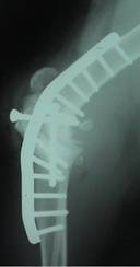
Hock (ankle) – Arthrodesis (fusion) is very effective at eliminating pain from the joint (see X-ray). Although it is technically more difficult to achieve than in the carpus the results are usually very good. As with the wrist, the commonest complications seen are infection and the plate used to “fix” the joint will need to be removed in about 25% of cases once fusion has been achieved, because of irritation under the skin.

In conclusion, therefore, osteoarthritis is a degenerative condition of a joint which cannot be cured. However, by adopting a sensible approach to management it is usually possible to return a dog to a good quality of active li
