Intervertebral Disc Disease / Rupture
What is an Intervertebral Disc?

The “back bone” of a dog is made up of a number of bones (the vertebrae) and between each pair of bones is an intervertebral disc. Each disc is made up of a jelly-like centre wrapped in a network of fibrous strands that blend into the bones next to the disc. This creates a flexible junction between each pair of bones and, along with the small joints between the vertebrae this allows movement between each pair of bones. Although this movement is fairly limited, because there are so many bones along the spine the amount of movement along the whole length of the “back bone” is quite large.
What Happens in Disc Disease and why does it Rupture?
The discs are present all along the spine but the areas where disc disease / rupture are seen tends to be in the areas where there is greatest natural movement: in the neck (discussed here and under “Wobbler Syndrome”); in the middle of the back, where the rib cage ends; and close to where the spine joins in to the pelvis (see under “Lumbosacral Disease”). When discs degenerate they dehydrate, that is to say they lose their water content. As a result the jelly in the centre dries out and becomes stiffer, in fact in the dogs with short legs it can become very chalky because of calcium being deposited in the tissue (which makes it visible as a white shadow on an X-ray – see later). The fibres
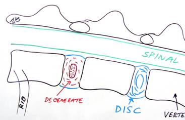
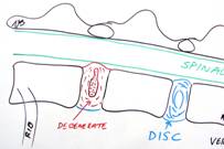
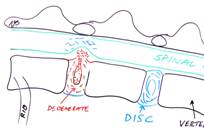
around the centre of the disc also dehydrate and break up, making them less effective at “containing” the core of the disc. The normal pressures on the disc as the dog moves can then cause the disc to bulge upwards as the material in the core pushes between the fibres. If that material breaks right through the shell of the disc then the disc ruptures and the material from the centre of the disc spills out into the vertebral canal. This causes damage to the spinal cord in two ways: the impact of the material on the spine causes bruising; and the mass of material in the canal causes pressure on the spine. It is the combination of bruising (contusion) and compression that results in the signs seen from the outside in terms of pain, paralysis etc
What Signs will a Dog show if they are suffering with this problem?
A dog with disc disease alone (i.e. no bulging or rupture) might show no signs at all. In fact in a study involving taking X-rays of clinically normal Miniature Dachshunds over two years of age, a high proportion (something like 70%) showed evidence of disc disease, though none showed any signs of the problem in terms of pain etc.
Signs relating to disc disease (but without any bulging or rupture of a disc) that a dog might show would relate to pain. In the case of such a problem in the neck they might show:
- Pain on moving their head suddenly
- Difficulty putting their head down to eat or drink
- Sudden acute onset inability to move, standing with their heads down and neck “fixed”
- When in pain they often “look with their eyes” rather than their heads – shuffling around to look at something rather than turning their head

Cocker Spaniel with severe neck pain caused by a ruptured disc
– holding his head down in this position all the time
In the case of such a problem in their back they might show:
- A tendency to walk with an arched back
- Difficulty / reluctance to jump up on furniture or small steps
- A tendency to cry out when picked up
- Less activity with more time spent in their bed
Signs relating to a disc rupture that a dog might show would relate not only to pain (as above) but might also include signs resulting from the direct injury to the spinal cord. In the case of such a problem in their neck they might show:
- Weakness / stumbling on a leg, most often one front leg
- Inability to stand and walk (paresis) – but sometimes a dog will not get up because of the severity of pain
- Inability to move any of their leg (paralysis)
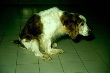
Springer Spaniel with hindleg paralysis caused by a ruptured disc in his back
In the case of such a problem in their back they might show:
- Weakness / stumbling on one or both hindlegs
- Inability to stand and walk on their hindlegs
- Inability to move their hindlegs
- Inability to control going to the toilet
How can it be proved that Disc Disease / Rupture is the cause of those Signs?
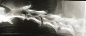

From the clinical history and signs being shown it is likely your veterinary Surgeon would have a very high index of suspicion that your pet is suffering with a disc problem. Standard X-rays can be helpful in showing evidence of degeneration, especially in the short-legged breeds, as their degenerate discs will often become white on an X-ray. However, it must be remembered that not all degenerate discs cause pain and so in many cases more than one disc is degenerate on an X-ray but which one is causing the signs would remain uncertain.
In dogs with a ruptured disc, standard X-rays may be helpful but are not entirely reliable. They might show evidence of disc disease and in some cases the space between the vertebrae is reduced where a disc has ruptured. However, this narrowing is not entirely reliable because it can be affected by how the patient is positioned for the X-ray and also cannot differentiate an old problem from a new one (unless previous X-rays have been taken and can be compared).
To show the presence of a disc rupture causing pressure on the spinal cord more advanced imaging is required. Currently the “gold standard” for evaluating spinal injury (including damage caused by disc rupture) is considered to be MRI. However, MRI scanners are not available on a daily basis in South Wales and, in fact, the current availability of high resolution scanners 5 days a week is restricted to centres mainly in the south-east of England. Fortunately, for dog’s suffering with acute disc ruptures, sufficient information for diagnosis and treatment planning can be gained from a technique called myelography. This involves the taking of X-rays after a contrast agent (dye) has been injected around the spine. This agent appears white on X-rays and so outlines the spinal cord showing any swelling or compression of this structure. From this it is generally possible to diagnose the cause of the problem as being a disc rupture and also to identify which disc has ruptured. Although this technique does carry a small risk to the patient, its advantage is that it only requires access to a standard X-ray machine and someone familiar with the technique itself. For this reason it remains the most commonly used procedure at Weighbridge Referral Centre for diagnosing disc problems in acute cases. However, we do arrange for dogs with less urgent conditions to have MRI scans “off site” (see under “Wobbler” or “Lumbosacral” syndrome).

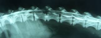
Plain X-ray – After myelography – the lines of dye are very thin over
shows one of discs is very white that disc confirming it has ruptured
How can We help these patients? – Surgery
Although dogs showing signs of disc disease can be managed “conservatively” (meaning cage rest and any required nursing) the results are less good than if surgery is undertaken (see below). If the “conservative” path is chosen and there is no improvement over several days then any thoughts on re-considering surgery have to accept a reduction in success rate caused by the delay. Furthermore, if signs are mild then acute deterioration might be seen with “conservative” management, which would then reduce the likelihood of recovery or compromise the quality of the recovery.
Surgery has, generally speaking, three possible goals in managing disc disease: to remove any ruptured disc material so that it is no longer compressing the spinal cord; to remove any remaining disc material from the core of that disc so that it is unlikely to rupture again, in the near or distant future; and to remove the core of neighbouring (other “high risk”) discs to reduce the likelihood of them causing problems in the future. It may not be possible (or necessary) to achieve all three goals in any particular patient owing to the nature of their disc disease, which disc is involved, breed of dog involved.

If pain is the only clinical sign being noted then it may be that the disc has not yet ruptured, in which case there might not be any compression of the spinal cord. However, it is then difficult to be sure as to which disc is actually responsible for the pain. As a result, a surgical technique called “fenestration” might be chosen. This involves creating a small window in the floor (neck discs) or side (back discs) of each of several (the “high risk”) discs and removing the core of each of these discs. This procedure is very effective in resolving pain as long as it is being caused by one of the discs included in the surgery – fortunately this is the case in the vast majority of dogs. It is also very good at preventing any serious recurrence of problems in the future since all the “high risk” discs have been dealt with at the same time.
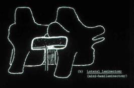
In cases with severe pain (neck discs) or poor mobility/paralysis then compression of the spinal cord is likely to be an issue and surgery needs to be targeted at removing that compression. In order to do this it is necessary to remove some of the bone from the vertebrae on each side of the affected disc to allow access to the vertebral canal so that the ruptured disc material can be removed (this is called a “ventral slot” procedure in the neck and a “laminectomy” in the back). Removal of the remaining “core” material from the affected disc is generally carried out at the same time. It might also be possible to “fenestrate” neighbouring disc spaces, which reduces the likelihood of future recurrence.
After surgery, particularly in those cases with obvious mobility issues, a programme of physiotherapy / rehabilitation is strongly recommended and, through collaboration with the SMART clinic, such a programme is generally started the day after surgery and then continued on an outpatient basis with that clinic once they have been discharged from Weighbridge Referral Centre.
How Successful is treatment of this problem?
How successful treatment of this condition is depends on a number of factors, including:
- Severity of the clinical signs
- Duration of the clinical signs – though this has most influence on the rate of expected recovery rather than the likelihood of recovery, except with very severe clinical signs
- Ability to nurse the patient and the patient’s co-operation with being nursed
- Veterinary Surgeon’s experience (if surgery is involved)
- Veterinary Physiotherapist’s experience (if physiotherapy is involved)
At Weighbridge Referral Centre, we regularly see dogs suffering with degenerative disc disease / rupture. The following comments regarding outcome are based on the results seen by Steve Butterworth over the past twenty years. It is important to bear in mind that an unsatisfactory outcome could mean there is no improvement in the clinical signs but it could also mean that there is a sudden deterioration.
In the majority of dogs showing signs relating to pain +/- mild weakness in one or more legs, the problem can be successfully managed (whether by conservative management or a need for surgery) in the vast majority of cases, though pain might take a few weeks to resolve completely.
In those showing marked weakness of two or more legs the results for a return to satisfactory quality function are good with conservative management (about 70%) but better with surgery (about 90-95%).
In those that are paralysed but still show they have feeling in the affected feet the results are fair with conservative management (about 50%) and, again, better with surgery (about 85-90%).
In those unfortunate patients that have lost feeling in the feet of paralysed legs the results are poor with conservative management (< 10%) and still only fair with surgery (50-60%).
Another aspect of long-term success is the likelihood of recurrence. This is far more common when disc disease affects the back rather than the neck. In either case the likelihood of significant recurrence following conservative management is about 30% within two years. Following surgery the risk is much less – somewhere between 1% and 15% depending on the nature of the surgery, disc involved in the fist episode, breed of dog involved.
Note on disc disease in Cats
Although disc disease does occur in cats it is rarely a cause of clinical signs. If signs like those described above do develop in a cat and the cause is proven to be a disc-related problem then treatment along the same lines as detailed above could be considered.
