Osteochondritis Dissecans (OCD)
What is OCD?
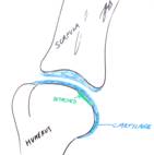
Osteochondritis Dissecans (OCD) is one form of a group of diseases called Osteochondrosis all of which relate to some sort of problem with growing cartilage being converted into bone. In the case of OCD the cartilage involved is that on the surface of a joint (or joints). Within any given joint the area of cartilage affected is fairly consistent and might relate to potential pressure points created in the joint by slight conformational (shape) abnormalities. The following explanation of how OCD develops is very simplistic but, hopefully, it will help an owner to understand the problem a little better. Because the growing cartilage on the surface of the bone within a joint fails to be converted into bone it become thicker. As it does so the deeper layers of the cartilage cannot gain sufficient nutrition and the tissue dies and loses its connection to the underlying bone (ii) below. A small defect develops underneath this area of cartilage. As weight is taken on the overlying cartilage it tends to “bounce” because of this defect and this can lead on to a crack developing at the margin of the underlying defect (iii) below. If this happens all the way around then the flap of cartilage comes away and may fall out of the way into a pouch of the joint capsule. From here it might be reabsorbed or it might persist as a so-called “joint mouse” (iv) below. Once the cartilage flap has fallen out of the way the defect tends to fill in with fibrocartilage, which although not as good as the original articular cartilage will usually work adequately well. However, problems arise when the cartilage flap does not come off and sits between the joint surfaces, much like a “pebble in a shoe”.
Schematic explanation for OCD of the Shoulder joint:


(i) (ii) (iii) (iv)
There is little doubt that the cause of OCD is, in part, heritable as the problem does tend to run in certain breeds and families. Also, which joint is affected tends to be breed related. OCD of the shoulder tends to be seen in large breeds such as Great Danes and Irish Wolfhounds but also in Border Collies, whilst elbow OCD is mainly seen in the Labrador retriever.
What Signs will a Dog with OCD Show?
It is important to be aware that not ALL dogs with osteochondrosis or OCD will show clinical signs and, in particular, may not show signs in both limbs despite the problem being bilateral (as judged from X-rays). If they do show signs then these will relate to lameness and obviously whether this affects a front or back leg depends on which joint is involved. In most cases of OCD the lameness will be seen in young dogs, less than a year of age.
If the signs are restricted to only one forelimb then the most obvious feature is the dog showing a nodding of the head as they walk or a tendency to lift or rest the paw when sitting (so as to keep weight off the limb). The nodding is a way of keeping the weight of their head on their good leg so it is the leg that is taking weight when the head is UP that is the bad leg. If the signs are present in both forelimbs then lameness might not be so dramatic because both legs hurt equally. As a result the signs relate more to showing a shuffling gait, reluctance to jump down or come down stairs, or simply a reduced willingness to exercise.
If the signs are related to pain in one hindlimb then the dogs may show a tendency to skip or miss a stride on that leg or stand with less weight on that foot. If both hindlimbs are affected then, in a similar way to what is seen in the forelimbs, they may show a shuffling gait, tendency to “bunny hop”, difficulty jumping / climbing stairs or a reduced willingness to exercise.
Whichever joint or joints are affected, another sign that may be seen is stiffness after rest following exercise. This sign is often related to arthritis that develops as a consequence of the irritation caused to the joint by the OCD.
How is OCD Diagnosed?
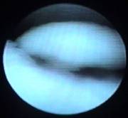
Your Veterinary Surgeon will probably have a strong suspicion that this is the problem in a dog of the “right” breed and age showing lameness associated with joint pain. Confirmation can usually be gained from X-rays that will show a defect in the bone underlying the affected area of cartilage. If this diagnosis is suspected but no such radiographic changes are present then it may be possible to show the presence of an OCD flap by a technique called arthrography (where a dye that looks white on X-ray is injected into the joint and this surrounds the flap and gets under the surface of the cartilage) or by arthroscopy (“magic eye”) where a small camera is introduced into the joint so that the cartilage surface can be seen. The images below are of OCD affecting the shoulder: (i) is an Xray of normal (on the left) and OCD affected (on the right) shoulders showing the defect in the line of the bone; (ii) is after arthrography where the dye can be seen running under the cartilage where the bone defect can be seen, indicating a cartilage flap is present; (iii) is a view of an OCD flap seen through an arthroscope.


(i) (ii) (iii)
How can OCD be Treated?
Conservative Management
In some cases showing signs OCD (of any joint) the signs will improve over a 4-6 week period of nothing more than controlled exercise +/- painkillers (if required). This may be a result of the cartilage flap becoming fully detached and either being resorbed or dropping into a pouch of the joint where it causes no problems. The defect in the cartilage can then heal by forming fibrocartilage and the signs might well resolve.
If patients are not significantly better after a 4-6 week period then surgical treatment should be considered.
Surgery
In general the surgery is targeted at removal of the cartilage flap and this can be done by open surgery or, in some cases, using “key hole” surgery with a “magic eye” or arthroscope. The recovery rate is a little faster if the flap is removed arthroscopically but the outcome is more related to the joint involved. With shoulder OCD the vast majority (over 90%) will be sound within 6-8 weeks of surgery and there are very few where lameness recurs at a later age because of arthritis. In the case of the elbow and hock (ankle) a satisfactory recovery will generally be seen in about 75% but many will show lameness associated with arthritis later in life. The stifle (knee) joint is not affected with OCD very often and the results of surgery can be quite variable.
Recent developments in surgery have looked at transplanting a plug of joint cartilage with its underlying bone to replace the cartilage that has been lost at the site of the OCD. The cartilage is taken from a part of the joint that is not used for weight bearing. Early clinical results using these techniques are encouraging and might help us to improve the success rates seen with the treatment of elbow and stifle (knee) OCD. The technique would be difficult to apply to the hock (ankle) and of little use in the shoulder where the results are already very good.
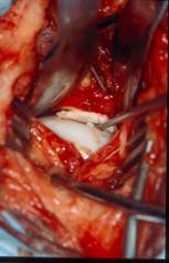
The images below show: (i) an OCD lesion of the shoulder as seen at surgery with the probe lifting the cartilage flap off the underlying bone; (ii) an OCD flap removed from a shoulder joint (iii) arthroscopy of an elbow joint with forceps grabbing onto an OCD flap.


(i) (ii) (iii)
Post-Surgery
After any of the surgeries mentioned above it is important that the patient is rested whilst healing takes place. Exercise is restricted to lead exercise of distances and frequency that does not cause significant worsening of the lameness and such that it can be repeated or slightly increased day by day. The recovery period is generally about 2 months. Physiotherapy / post-operative rehabilitation can be useful in improving the rate of recovery.
What will happen in Later Life if a Dog is affected by OCD as a Puppy?
If a puppy has had OCD, whether or not they have shown any clinical signs at that age and irrespective of how they were treated, in later life they might develop signs associated with secondary osteoarthritis (OA). Such signs might involve stiffness after rest, lameness or poor exercise tolerance. The likelihood of developing such signs is related to which joint has been affected – involvement of the elbow or hock (ankle) is far more likely to result in OA at a later age than if the shoulder or stifle (knee) had been affected. Further information on managing OA can be found under “Osteoarthritis”.
